
Princeton University举办的Art
of Science Competition

In the spring of 2006 we again asked the Princeton University community to submit images―and, for the first time, videos and sounds―produced in the course of research or incorporating tools and concepts from science. Out of nearly 150 entries from 16 departments, we selected 56 works to appear in the 2006 Art of Science exhibition.
The practices of science and art both involve the single-minded pursuit of those moments of discovery when what one perceives suddenly becomes more than the sum of its parts. Each piece in this exhibition is, in its own way, a record of such a moment. They range from the image that validates years of research, to the epiphany of beauty in the trash after a long day at the lab, to a painter's meditation on the meaning of biological life.
We thank all those who submitted their work to this year's competition. By sharing their imagination, as well as the fruits of their research, they have reaffirmed the deep links between art and science.
.
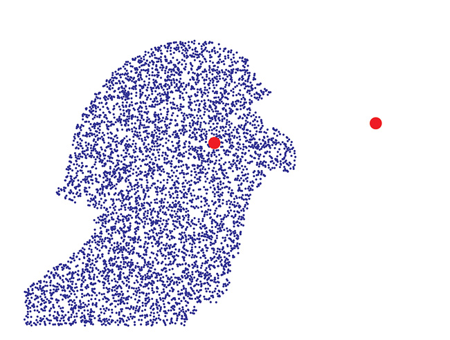
One or the Other
Darren Rand GS, Jayson Paulose ‘07, and Ken Steiglitz
Departments of Electrical Engineering, Physics, and Computer Science
This plot was generated from a model describing collisions of optical
solitons, beams of light which do not diffract. Pictured are two basins
of attraction, given by the white and blue regions, along with the corresponding
foci (red dots), plotted on the complex plane.
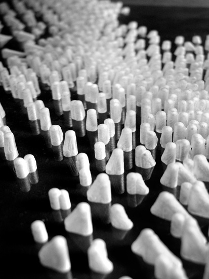
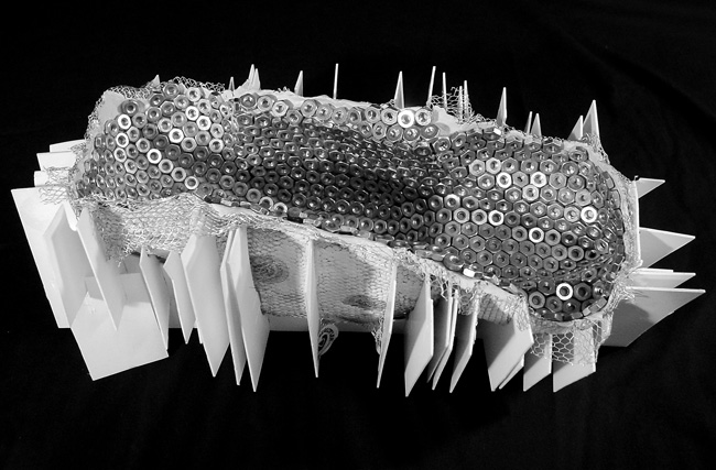
Mapping Urban Fluxes
Aurel von Richthofen GS and Hyundai Kim GS
School of Architecture
Preliminary to the design of a fashion institute in Milan the authors,
two students at the School of Architecture, tracked vectors of horizontal
disruption as interpretation of complex urban fluxes. While vertical vectors
of gravity forces inform the structure of many buildings and find their
architectural expression in columns, beams and trusses, horizontal vectors
such as vectors of movement and sight, can only be simulated using software
deriving from the animation industry. A fluid dynamic simulation within
the 3D model of the site generated patterns of density. These were then
exported into computer aided design software that interpreted the patterns
as fields of spatial vectors.
In order to reinform the design project for the fashion institute with
the vectors of horizontal disruption found through the fluid dynamic simulation
a physical model of the field was needed. The laboratory at the School
of Architecture is equipped with some of the latest computer-driven fabrication
technology for model making, including a Precix 9100 series large-bed
milling machine. The machine consists of a router and a 3-axis mill over
a large table that drives a spinning bit into materials such as acrylic,
wood or metal. Code generated from the virtual model drives the mill.
We used
the machine to produce a series of 12 by 12 inch acrylic models from which
the pictures shown are taken. The models were produced upside down, flipping
the milled side to the bottom and producing a negative image of the field.
The diameter of the milling bit imprints the vectors as fins into the
acrylic. The transparency of the material and the refraction of the light
create a haptic impression even though the vectors are captured inside
the material.

Desert Jewels
David Potere GS
Office of Population Research
This image of irrigated agriculture in the deserts of central Saudi
Arabia, 450 km west of Riyadh, was taken by the Landsat 7 satellite on
February 5, 2000, while orbiting 700 km above the surface of the Earth
at a speed of roughly 26,000 km/h. The Saudis manage to make the desert
bloom by pumping fossil water from deep below the Earth’s surface. A well
at the center of each of these fields feeds a center pivot irrigation
system which spreads water in large circles up to one kilometer in diameter.
The aquifers which supply these fields are ancient and finite. When the
fossil water runs out, the desert sands will return. Like the irrigation
projects of many arid regions, the Saudis’ desert jewels will soon fade.
I began building this image by selecting a set of infrared bands that would best tell the story of irrigated agriculture in Saudi Arabia. Landsat satellites see the earth through eight spectral bands―the reds, greens, and blues of human experience, along with much longer wavelengths of infrared and thermal light. After using image processing software to assemble the initial false color composite, I selected a 100km-wide subset and choose contrast and saturation levels to accentuate the most interesting features of the image. Because healthy vegetation reflects strongly in the near infrared, the Saudis’ alfalfa and wheat fields are painted red against the desert background. To an astronaut these fields would appear green, and the intense brightness of the desert would likely washout the complex mineralogical patterns picked up by Landsat.
Aside from
providing beautiful images, Landsat is used in desert environments to
build maps of irrigated agriculture, to prospect for water and minerals,
and to manage natural resources. The workhorse of NASA’s Earth observing
satellite constellation, Landsat-series satellites have been on orbit
continuously since 1972, making Landsat the longest-running satellite
collection program in the world.
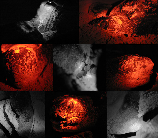
Nesting
Leatherback Sea Turtle
Celene Chang ‘06
Department of Ecology and Evolutionary Biology
Once categorized with mythical sea creatures, the leatherback sea turtle
(Dermochelys coriacea) is perhaps the most majestic reptile alive. Rather
than a typical hard shell, it is covered with leather-like skin, optimal
for deep-diving and withstanding cold temperatures. Not only is it the
largest of the sea turtles (up to 70 inches in length), it is the fastest
moving and deepest diving: it can dive to 1230 meters for food, the same
depths as a whale. These photographs were taken during the most extraordinary
time of the adult turtle’s life: nesting. The nesting female is in a state
of complete hypnosis. This trance-like state, along with her blindness
to red light, allowed us to stroke her body and head, and witness the
egg-laying from point blank. After slowly emerging from the ocean, she
dug a deep cavity with her rear flippers. She then laid her eggs, visibly
straining in each contraction through her Lamaze-style breathing. When
she finished, she masterfully covered the nest until its plot was indistinguishable.
Although the tears in the center right photograph were mucosal extract
to moisten her air-exposed eyes, it was difficult not to attribute them
to pain and utter exhaustion. Classified as a critically endangered
species, this female is presumed to be one of only 200 turtles that nested
on that Panamanian beach in 2005. Undoubtedly the dinosaur among the seven
sea turtle species, creatures such as this leatherback are powerful reminders
of the magnificent megafauna that walked the earth millions of years ago.

Interior Vacuum Vessel NSTX
Elle Starkman and Charles Skinner
Princeton Plasma Physics Laboratory
The National Spherical Torus Experiment (NSTX) is an innovative magnetic
fusion device that was constructed by the Princeton Plasma Physics Laboratory
(PPPL) in collaboration with Oak Ridge National Laboratory, Columbia University,
and the University of Washington at Seattle. This image is of the interior
of the experiment showing the protective carbon tiles and the central
column. Various diagnostics are mounted at the midplane.
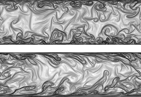
Turbulent Channel Structures
Melissa Green
Department of Mechanical and Aerospace Engineering
Turbulence is chaotic by definition, but previous work has shown that
turbulent flow is organized into coherent structures. We use a method
called Direct Lyapunov Exponent to identify these structures in a turbulent
channel flow. The top image is a plane seen by looking down the channel,
and the bottom image is a plane seen from looking from the side of the
channel. Structures are outlined in black, and are more dominant near
the walls, as expected.
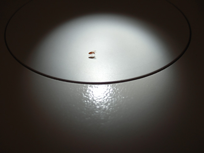
The Fly Show
Matthieu Coppey
Lewis Sigler Institute for Integrative Genomics
A mutant fruit fly Drosophila Melanogaster, shot under the light of
a Zeiss Stemi 2000 microscope. During the last three decades, the fruit
fly has been a central model for the study of development. A great number
of genetic modifications help the scientist in his discoveries, mainly
by linking the mutation of a gene of interest with easy-to-recognize phenotypic
attribute. Here, as a main actor of the actual science, a mutant with
curly wings and white eyes shows up...
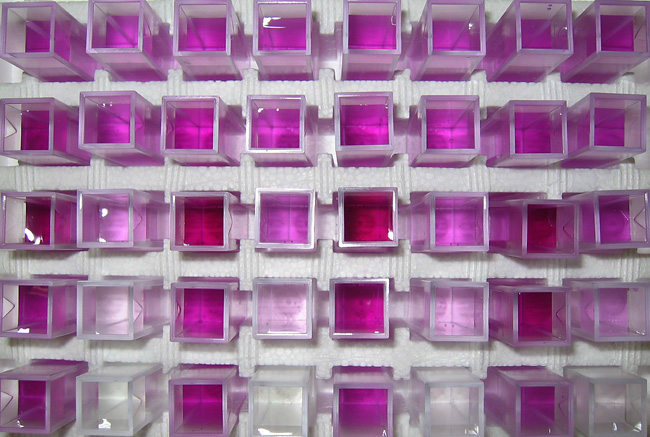
Color Patterns of an Iron Extraction Time Series
Andrew Altevogt
Department of Civil and Environmental Engineering
Hydrochloric acid flows in a closed loop through a column packed with
a synthetic iron oxide coated sand. As the acid contacts the sand it extracts
Fe(III) from the sand. The iron in solution causes the ferozine to turn
shades of pink/purple (darker shades indicate higher iron concentrations).
A spectrophotometer measures the absorbance of a specific wavelength of
light for each sample which is then correlated to iron concentrations.
Going from left to right and from top to bottom are a time series of samples
taken from the initial time until the equilibrium time when all iron has
been desorbed. Each set of two samples (left to right) are the outflow
(initially pink) and inflow (initially clear) from the column. The outflow
samples start relatively high (pink) become higher (more purple) and then
gradually become lighter (pink) again. The inflow samples start out clear
and gradually become darker (pink) throughout the experiment. The difference
between each pair of inflow and outflow colors is a representation of
the iron extraction rate. The extraction rate is high (clear inflow versus
pink/purple outflow) at early times and goes to zero, when all of the
iron has been extracted, at the end of the experiment (solutions are the
same shade of pink). The color patterns which arise are simply a manifestation
of dynamic changes in iron extraction.
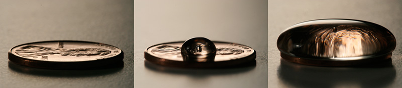
Tension
Ashwin C. Atre ‘09
Department of Chemical Engineering
Partial charges of hydrogen bonding give water its remarkable sticky
and elastic properties on display. The surface tension of the water droplet
above the coin shapes a seemingly impossible, yet beautiful and almost
magical form. The droplet clings on to the edges of the metal until the
intermolecular forces are no longer strong enough to resist the force
of gravity.
Lounge Chair (Image 4)
Emmet Truxes ‘06
School of Architecture
This project became an unintentional study of light, specifically the
reflective, refractive, and transparent properties of the plexiglass modules.
Due to the hundreds of angles of the normals, there is no telling which
modules will reflect the artificial light from ceiling and wall fixtures
or natural light from windows. When walking by the chair, the viewer is
able to see the lights bouncing to different modules, adding a distinct
level of animation to the experience. Furthermore, any reading is dependent
on the viewer’s height, his figurative relationship to the frame of the
piece, and the levels of interior and exterior lighting. These factors
drive how light is perceived by the viewer and ensure that no two readings
will be the same.
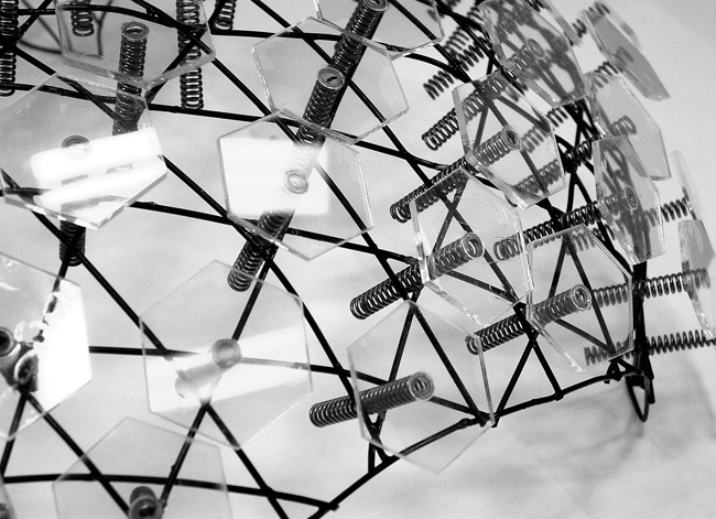
Lounge Chair (Image 1)
Emmet Truxes ‘06
School of Architecture
The formal and theoretical inspiration for this chair prototype results
from projects produced in the first half of Jesse Reiser’s spring 2005
undergraduate design studio in the School of Architecture at Princeton
University. The last part of a comprehensive study of surfacing techniques
focuses on the single modular unit as skin. The regular hexagon presents
itself as a suitable module because of its ability to self-tessellate.
Hundreds of hexagon nuts form the surface for the design model, acting
as planar modules surfacing a complex curved form. As the rows of hexagons
twist around the curves, they react and break from their neighbors, forming
negative space.

Cryptic Coalition
Trond H. Larsen GS
Department of Ecology and Evolutionary Biology
In addition to cryptic coloration allowing them to blend in with the
tree trunk, these Peruvian caterpillars fool their enemies by foraging
together in a large group. As a whole, the caterpillars may appear to
be a large patch of lichen. However, every individual must stay tightly
within the group in order to maintain the illusion. Moving in groups can
also have other benefits, including diluting attacks from predators and
parasitoids and increasing foraging efficiency through cooperation. This
provides one example of how simple interactions can scale up to form collective
behaviors that benefit the species. The rules of movement can be estimated
from the turning patterns of individuals in the photograph. Other species
use related strategies, such as moving in a single file line which may
be interpreted as a snake or a liana.
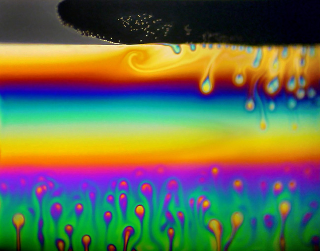
Soap Film Hurricane
Steffen Berg and Sandra Troian
Department of Chemical Engineering
The image shows white-light reflection from a flat soap film formed
from an aqueous solution of the anionic surfactant sodium dodecyl sulfate
and the hydrosoluble polymer poly-(ethylene oxide). The planar film was
formed in a 10 mm wide, vertical frame. The very dark upper region of
the film, the so-called “black film” region, is less than 100 nanometers
thick and has low optical reflectivity. A step-wise thinning front in
the black film region progresses from right to left and causes a swirl
in the yellow soap film band. The peacock feather patterns in the yellow
and green parts of the film arise from inhomogeneous distributions of
the surfactant causing gradients in the surface tension.
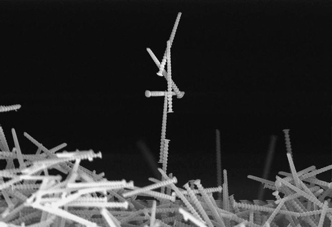
Above the Fray
Keith Morton GS
Department of Electrical Engineering
Scanning electron micrograph of 300nm diameter high-aspect ratio silicon
pillars made using nanoimprint lithography and deep reactive ion etching.
The original, regular array of pillars was part of a microfluidic device
to separate nanoparticles. The pillars pile up from scribing damage when
the silicon wafer is cleaved to obtain a cross-section image.

Fairies
Margaret E. Bisher and Soyeon Im
Department of Molecular Biology
The “fairies” in the image above occurred unexpectedly in a sample of
protein filaments. They were visible in addition to the expected rope-like
structures. The “fairies” showed up again a few weeks later, in other
protein filament samples. They are likely staining artifacts: a result
of the stain precipitating on the carbon film that supports the sample.
These odd structures form randomly and very intermittently. The original
protein filament sample itself was in a solution or buffer that was absorbed
onto a grid (a mesh-like structure 3mm in diameter) with a carbon support
film across it, approximately 50 Angstroms thick. The sample was washed
with water and then stained with 1% uranyl acetate. It was then viewed
at 80kV on a Zeiss 912AB transmission electron microscope equipped with
an Omega energy filter.

Trapped Terrapin
Madeline Renny ‘06
Department of Ecology and Evolutionary Biology
This image displays a female northern diamondback terrapin, Malaclemys
terrapin terrapin, captured in a crab trap in the salt marsh of southern
New Jersey. This terrapin was captured for research purposes for the Terrapin
Conservation Project at the Wetlands Institute in Stone Harbor, New Jersey
(this was also the research base for the field work for my thesis). However,
each year thousands of terrapins drown from being caught in crab traps
that do not have the proper excluder devices. The peaceful nature of the
terrapin and the reflection of the sunlight on its shell contrast with
the restraining bars of the crab trap.

Perturbed Circle of Life
David Karig
Department of Electrical Engineering
A twirling mesh of bright synthetic blue hands reaches out to me... Pictured
are the contents of a biohazardous waste bin in our synthetic biology
lab. Our research group focuses on engineering life and “programming”
living cells such as E. coli, yeast, and stem cells. I was captivated
by the colors in my delirium during a non-stop 36 hour experiment.

Infection
Miguel Gaspar GS
Department of Molecular Biology
Electron microscopy image of human cytomegalovirus-infected human foreskin
fibroblasts at 96 hours post-infection. Virions and dense bodies can be
observed in the cytoplasm. In collaboration with Peggy Bisher from the
Confocal/EM facility of the Molecular Biology Department.
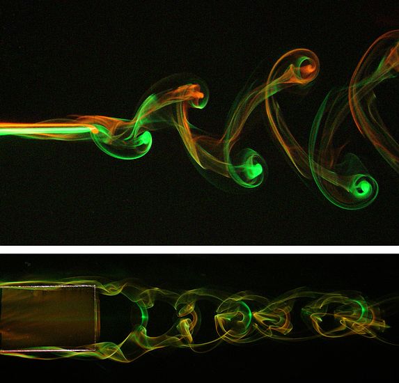
Wake of a Pitching Plate
James Buchholz GS and Alexander Smits
Department of Mechanical and Aerospace Engineering
These images contain top and side views of the wake produced by a rigid
plate pitching about its leading edge in a uniform flow (flowing left
to right). The leading edge of the plate is hinged to the trailing edge
of a stationary symmetric airfoil. The wake is visualized using fluorescent
dyes that are introduced through a series of holes on each side of the
airfoil support. Twice in each flapping cycle, a horseshoe-shaped vortex
is shed from the top, bottom, and trailing edges. The vortices become
entangled to form the chain-like structure shown here. Studying such wakes
is believed to be important for understanding the mechanisms of thrust
production in fish-like swimming.
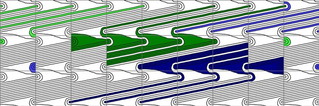
(19,3,12,3)
Gabriel Doyle '05
Department of Mathematics
This picture is a representation of the universal cover of the doubly-pointed
Heegaard diagram of genus 1 of a (1,1)-knot. The black line represents
the bounding curve for the knot, and the gray lines represent a meridian
and a longitude of the torus. By finding all disks bounded by a vertical
segment of the gray lines and any segment of the black lines, one is able
to calculate the Knot Floer Homology of a (1,1)-knot. This is a knot invariant
that can be used to tell similar knots apart. All 22 disks that can be
used to determine the Floer Homology of this knot are marked on this diagram,
with green and blue referring to the multiplicities of two special points.
Bright colors indicate disks with multiplicity 1, while the dark disks
have multiplicity 2. The title of the diagram is a set of four integers
that define this particular knot: the number of intersections between
the black and grey line on one side of the fundamental region (one small
square in the picture), the number of disks on one side of the fundamental
region, the number of lines going above the right-hand side’s disks, and
a rotation number.

Knowledge is Beautiful
Elyse Graham
'07
Department of English, Comparative Literature studies
This image is the first from a series of 12. They depict some of history's great scientific minds with the seductive physical draw that their minds hold for us intellectually. It's a laugh, but it's also a recollection of Blaise Pascal's "Clarity of mind is clarity of passion." We practice science because we love it. So, too, do we practice art. The figures are given male heads and female bodies in part to appropriate the old Platonic notion that divides "Mother" Nature (wild, sensual, unpredictable) and "Father" Science (logic and linearity). Contemporary investigation, criticism, and mixing between different sciences and spheres (History of Science, astrobiology, and so on) is opening those borders to new fields of analysis. The imagery also interrogates the traditional male-centeredness of science, and looks to an opening world in which science is performed by, and for, all of humanity rather than a restricted category or sex